Let’s Ruminate on it
Ruminants are artiodactyl characterised by their possession
of stomachs with four compartments. They are said to have four “stomachs” which
is technically untrue as they have only one stomach but divided into four
parts: the rumen, reticulum, omasum and abomasum. Nearly 200 wild ruminant
species exist including cows, goats, deer, giraffes, moose and elk.

Origin of the
Ruminants Stomach
When the first ruminant groups emerged, they were rabbit
sized (< 5kg; Hackmann & Spain, 2010). Their skull and dental morphology
were optimal for consuming fruits and insects. This shows that the first
ruminants were small, forest welling omnivores (Webb, 1998b). The first
ruminants did not have a functional rumen until about 40ma as indicated by
dental morphology and molecular techniques (Hackmann & Spain, 2010). At about
18 to 23 ma, new families of ruminants evolved with these families having masses
of 20kg to 40kg. The new families ate primarily leaves as evidenced by dental
wear (Hackmann & Spain, 2010). By about 5 to 11 ma, grasslands had expanded
and some species began including more grass in their diets leading to the
development of more complex stomach to help in the digestion of the grasses.
Structure and
Histology of the Ruminant’s Stomach
The ruminant’s stomach has four compartments: the rumen,
reticulum, omasum and abomasum.
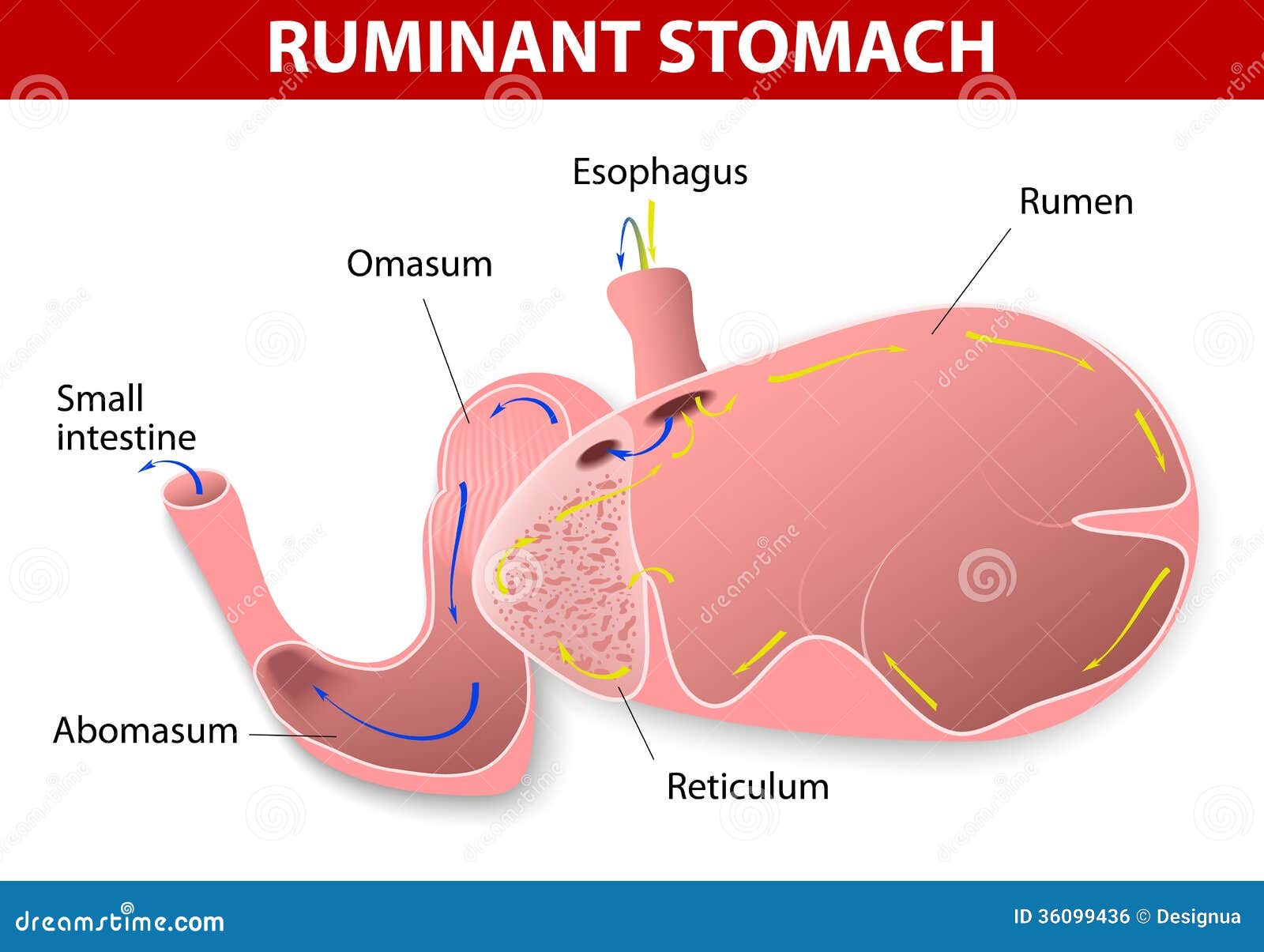
 Rumen: this is
the first and largest compartment of the stomach. After a ruminant feeds, food initially
collects in the rumen. The rumen is saclike, thin walled and lined and lined
with numerous projecting papillae that increases its absorptive surface area. The
rumen has highly vascularized connective tissue covered by stratified squamous
epithelium. The epithelium is involved in the absorption of short-chain fatty
acids (i.e. sodium salts, the microbial breakdown products of cellulose). This
transepithelial transport is highest in areas where the cells appear to be
swollen (Kwan, P.). Muscularis mucosae are absent in this part of the
forestomach. The muscularis external contains two layers of smooth muscle
fibers. Myenteric plexus can be found between
Rumen: this is
the first and largest compartment of the stomach. After a ruminant feeds, food initially
collects in the rumen. The rumen is saclike, thin walled and lined and lined
with numerous projecting papillae that increases its absorptive surface area. The
rumen has highly vascularized connective tissue covered by stratified squamous
epithelium. The epithelium is involved in the absorption of short-chain fatty
acids (i.e. sodium salts, the microbial breakdown products of cellulose). This
transepithelial transport is highest in areas where the cells appear to be
swollen (Kwan, P.). Muscularis mucosae are absent in this part of the
forestomach. The muscularis external contains two layers of smooth muscle
fibers. Myenteric plexus can be found between
Reticulum: this
is the most cranial part of the stomach and food and sometimes objects drops in
here first. The reticulum consists of mucosa, submucosa, tunica muscularis and
serosa. The reticular walls are lined with mucous membranes that are organized
in folds (laminae) to form a honeycomb pattern. On the laminae are short
conical projections called papillae. The lamina epithelial is keratinized
stratified squamous epithelim. Contractions of the honeycomb cells, with the
purse-string action of the smooth muscle strands, help the mechanical digestion
of the ingesta. The reticulum contracts to slosh the ingesta between itself and
the rumen (Kardong, 2002).

Omasum: this is
the third portion of the system that ingesta pass through in the ruminant. It
is spherical in shape and located to the right of the rumen and reticulum, as
well as being the smallest of the four compartments. The inside walls are
covered in muscular laminae that are studded with short papillae. The mucosa
and portions of muscularis mucosae form parallel folds that resemble pages of
an open book. The mucosal surface is covered by stratified squamous epithelium.
The core of the laminae are quite characteristic with three layers of smooth
muscle. Extending upward from the muscularis mucosae are fibers that form the
two outer layers. The orientation of the muscle cells is parallel to the free
edge of the laminae. Sandwiched between these two layers is a single inner
layer of smooth muscle. This is derived from the muscularis externa and the
fibers are perpendicular to those of the outer layers. The omasum operates like
a two phase machine. First, there is relaxation of the muscular walls which aspirates
fluids and fine particles from reticulum into the omasum (Kardong, 2002). Second,
the omasum contracts to force the digesta into the abomasum. The omasum absorbs
volatile fatty acids, ammonia and water and at the same time separates the
fermenting content of the rumen and reticulum from the highly acidic content of
the abomasum (Kardong, 2002).

Abomasum: this is
the fundic part of the stomach in which further digestion occurs before digesta
pass to the intestine. It is the glandular portion of the ruminant stomach and
its histology is highly similar to that of monogastric animals. The lumen of
the glandular stomach is lined with simple columnar mucus-secreting epithelium. The mucosa is thrown up into longitudinal folds called rugae which stretch flat as the stomach
distends. The lamina propria is a loose connective tissue layer
rich in capillaries and lymphoid cells and is entirely occupied by glands, the gastric glands (Kardong, 2002). The
abomasum produces hydrochloric acid and digestive enzymes, such as pepsin
(breaks down proteins), and receives digestive enzymes secreted from the
pancreas, such as pancreatic lipase (breaks down fats). These secretions help
prepare proteins for absorption in the intestines. The pH in the abomasum
generally ranges from 3.5 to 4.0. The chief cells in the abomasum secrete
mucous to protect the abomasal wall from acid damage (Kardong, 2002).


Development
of the Ruminant Stomach
No comments:
Post a Comment